Vision Motion Sensitivity/Visual Vertigo
In this blog we will define Vision Motion Sensitivity (VMS), discuss the most common causes, review the current forms of remediation, and share some exciting new advances in the area.
What is Vision Motion Sensitivity?
Vision motion sensitivity (VMS), also commonly known as visual vertigo (VV), is a condition where strong visual stimuli can induce feelings of dizziness, vertigo, loss of balance, and even motion sickness resulting in nausea.
Our peripheral or magnocellular vision functions as our motion detector, providing information about the body’s movement through space and motion occurring within that space even when the body is not in motion. Our central vision gives us details about an object or scene once the eye muscles, with the help of the vestibular system, move the central or foveal vision to align on that object.
VMS or visual vertigo may occur because of a mismatch of information between the peripheral and central vision systems, a mismatch between the visual systems and proprioception, or most commonly when there is a conflict between the information from visual system and the vestibular system. Or more simply put, when the eyes are telling you one thing, and the inner ear is telling you another, it can get very confusing and overwhelming.
What are some of the causes for VMS/VV?
Neurological insult to any of the three systems (vision, vestibular, somatosensory) that control balance, or the nerve fibers that integrate information between them can result in VMS or VV. Common causes include traumatic brain injury from accidents or falls, concussions, strokes or other vascular incidents, and hypoxic events such as choking.
Traditional treatment for VMS
Unfortunately, immediately after a trauma or incident, most patients must deal with VMS through avoidance and adaptation. We have them avoid those situations or environments, such as a busy grocery store or driving on busy roads, or anything that would exacerbate the symptoms. These changes in lifestyles may often add to the subject’s anxiety and depression by taking away simple things that give them joy. Adaptive strategies such as limiting the time in the store, going at off peak times, reducing the extent of peripheral visual field with occlusion, and various tinted lenses, also are employed. Adaptation and avoidance should be the start of the journey for these patients, as it does give them time to heal, it should not be the only “therapy” utilized.
Traditional treatments for VMS involve vestibular rehabilitation with an emphasis on the vestibular-oculomotor reflex (VOR), such as VORx1 and VORx2 type therapies. Vision therapy techniques, especially for saccadic eye movements and binocularity, are often a critical need for many patients as well.
Even pharmacological treatments, especially acetazolamide, have been studied and show potential symptoms reductions for some patients. [1]
Many articles and blogs have been written to address these types of therapeutic protocols [3] so in this blog we will concentrate on newer therapeutic techniques that have arisen with the help of new advances in technology.
Using the latest technology to assist in VMS/VV therapy
The goal of many therapies is to promote desensitization to visual stimuli and increase tolerance. There are several different philosophies as to how this is best accomplished, with the two most common utilizing peripheral lock and central stimulation [2], and the other being the use of optic flow patterns that primarily move from central to peripheral. There is great merit in both philosophies, with many patients benefiting from a combination of both. Today we are fortunate to have the instrumentation and technologies that allow us to easily provide both these techniques to our patients, while being able control the speed, content, contrast, direction, and field of view of the visual stimulus.
The newest treatments for visual vertigo or VMS utilize various levels of immersive environments and controlled peripheral stimulations to desensitize patients to visual stimuli that creates many of their symptoms.
The BVT (Bertec Vision Trainer) and similar large screen devices can control the visual environment with a field of view (FOV) of approximately 80 degrees, depending on the screen size and distance to the screen. These devices are excellent starting points for VMS therapy because they are only partially immersive as they provide a strong peripheral vision lock which tends to anchor or ground the subject to his or her world. Various forms of optokinetic (OKN) and visual flow patterns are then presented to the subject while monitoring the effects on balance if a force plate is available and utilized. The complexity, field of view, direction, contrast, and speed of the presentation can all be controlled to tailor the therapy to the subject’s needs. Slow, central, simple, low contrast patterns are the least disturbing for most patients. As therapists, we always strive to start therapy at the point where the patient can demonstrate some measure of success, building skills and confidence as the therapy progresses. As patients demonstrate success, one or all of these variables may be incrementally made more difficult. Which variable to change first is the judgement of the therapist and often based on case history and symptoms. Therapy is often started with the subject in the seated position, advancing to standing, and then to standing on foam as the patient demonstrates the ability to advance without risk of falling. Multitasking or adding a cognitive demand during therapy may also be added in at any time, but if often saved till the end.
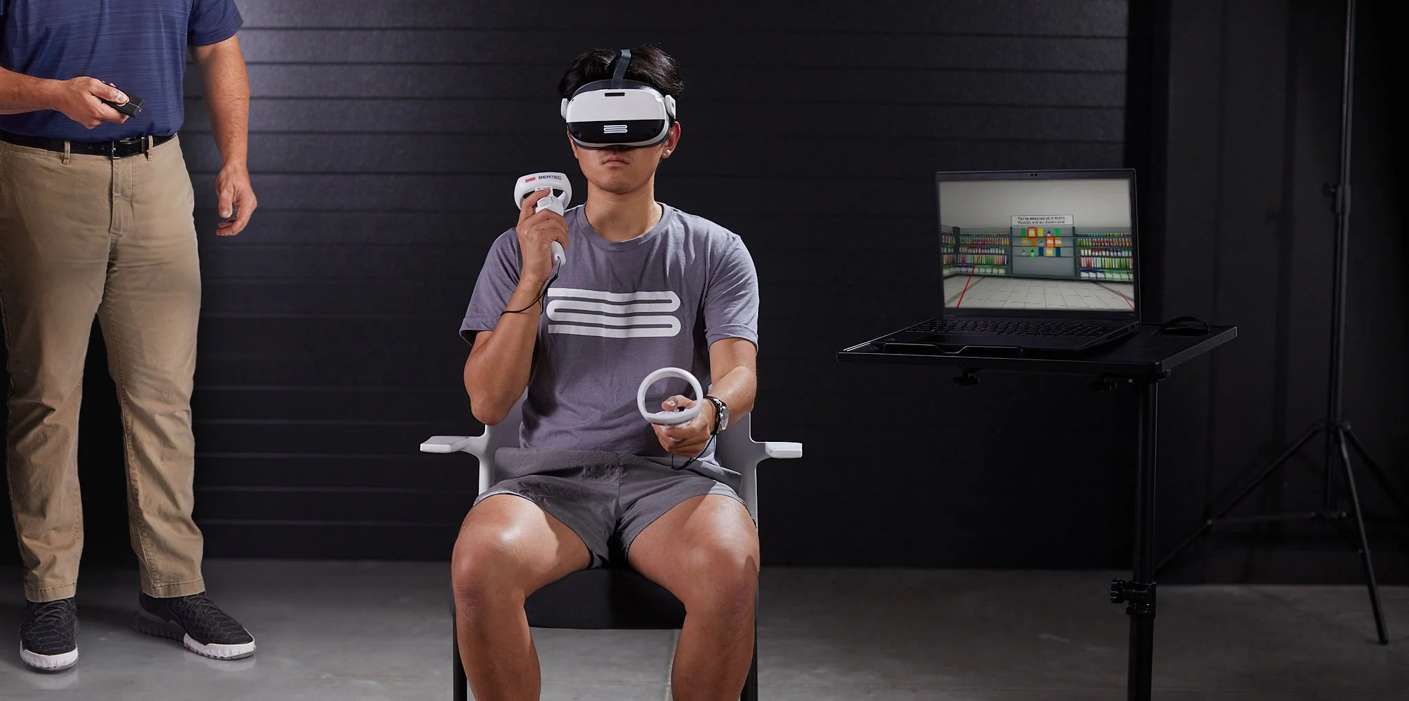
Another advancement in the treatment of VMS or VV is the head mounted display. HMD’s have a much higher degree of immersion than large screen interactive devices such as the BVT, as the only thing the subject sees is that which is displayed on the device’s two screens. The field of view is approximately 105 degrees for most HMD devices, somewhat less than you might think. This often seems larger because most HMD devices and their software create additional peripheral awareness by utilizing the technique of adding continuous peripheral views of the scene as the subject turns their head or moves it up and down. This gives the impression that the stimulus “wraps” totally around them. As with the BVT, the presentation of the stimulus can be very precisely controlled for speed, complexity, contrast, and direction. Additionally, the HMD can create true three-dimensional imaging through the binocular presentation of unique images to each of the two eyes. Patients with amblyopia or strabismus may not always be able to take advantage of this feature. This will also allow us to address binocular vision issues in the future as new software is developed. The HMD therapies, like the BVT therapies, should first be done seated, progressing to standing on firm surfaces and finally standing on foam to remove a portion of the somatosensory input, adding to the balance challenge. The clinician should always assess for safety and fall risk as the standing challenges are added.
The Computerized Dynamic Posturography device (CDP/IVR) is perhaps the ultimate device for both the assessment and treatment of visual vertigo and vision motion sensitivity. Although not a totally immersive device, it gives us the best of everything we are looking for. The field of view or presentation of stimuli extend past 180 degrees, encompassing the entire field of vision except for the feet and hands. This exposure of the feet and platform allows for some peripheral lock or grounding which reduces the distress many find when placed in a totally immersive visual reality environment with no obvious visual reference point. The CDP’s safety harness combined with the clinician’s demonstration and assurance the test or training may be stopped at any time reduces the risk of falling and helps reduce patient anxiety when in the system. Combined with the safety harness safety demonstration, the clinician’s reassurance reduces the risk of falling and the patient’s anxiety during assessment and training. This reduces risk of fall while in therapy, reduces patient anxiety while doing the tasks, and allows for better acceptance of the therapy protocols. Simply put, it is harder to concentrate on the therapy tasks you are given if you do not feel grounded in space and feel out of control, or that you may fall.
We can totally control the variables mentioned before such as contrast, speed of presentation, lighting, complexity of the scene, and cognitive tasking. Special visual flow scenes have been created to simulate walking in a park, walking down a grocery store aisle, driving, sidewalks and various optokinetic stimulation environments. These scenes were designed to control the complexity and speed of the simulation and add in other unique variables such as obstacles and cognitive demand.
In addition to all the techniques mentioned above, the platform the subject stands on is dynamic. The tilting or movement of the platform adds a whole new dimension to therapy, allowing for real world integration of posture and balance into the visual motion sensitivity therapy approach. This dynamic therapy should only be added after the patient has reached the appropriate milestones of VMS therapy on a stationary platform. The subject would ideally be the absence of all VMS symptoms while on a stationary platform and having little or no sensitivity to strong peripheral stimuli while walking on flat, non-patterned surface.
Visual vertigo or vision motion sensitivity has been one of the most difficult challenges we face as clinicians. This has been in part because of the nature of the problem and in part due to a lack of effective therapies to remediate this anomaly. With today's advances in interactive display technology across touchscreens, HMD’s, and VR domes, we can create and control an extremely immersive environment and most of the details within it. This in turn allows us to change the speed, contrast, and other details within the environment to gradually de-sensitize the symptoms and remediate the effects of this imbalance between the visual, vestibular, and somatosensory systems.
References
Sluch IM, Elliott MS, Dvorak J, Ding K, Farris BK. Acetazolamide: A New Treatment for Visual Vertigo. Neuro-ophthalmology. 2017 Aug 2;41(6):315-320. doi: 10.1080/01658107.2017.1326944. PMID: 29344071; PMCID: PMC5764056.
Chang, C.-P., & C. Hain, T. (2020). Visual vertigo treatment through optokinetic stimulation with stationary anchoring. Physiotherapy Research and Reports, 3(1). https://doi.org/10.15761/prr.1000133
Hall, C. D., Herdman, S. J., Whitney, S. L., Anson, E. R., Carender, W. J., Hoppes, C. W., Cass, S. P., Christy, J. B., Cohen, H. S., Fife, T. D., Furman, J. M., Shepard, N. T., Clendaniel, R. A., Dishman, J. D., Goebel, J. A., Meldrum, D., Ryan, C., Wallace, R. L., & Woodward, N. J. (2022). Vestibular Rehabilitation for Peripheral Vestibular Hypofunction: An Updated Clinical Practice Guideline From the Academy of Neurologic Physical Therapy of the American Physical Therapy Association. Journal of neurologic physical therapy: JNPT, 46(2), 118–177. https://doi.org/10.1097/NPT.0000000000000382
Han, B. I., Song, H. S., & Kim, J. S. (2011). Vestibular Rehabilitation Therapy: Review of Indications, Mechanisms, and Key Exercises. Journal of Clinical Neurology, 7(4), 184. https://doi.org/10.3988/jcn.2011.7.4.184